Stress Echo Test
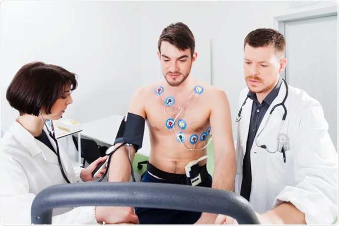
An exercise stress echo, sometimes simply called a stress echo, shows how your heart works when it is taxed. The test resembles a traditional exercise stress test. A technician will monitor your heart rate and rhythm as well as your blood pressure (this is standard during a stress test). But they will also use echo imaging (which is not normally used during a stress test).
This test shows how well your heart can withstand activity. Your sonographer takes pictures before you start exercising and then right after you are done.
In some cases, you will not exercise. Instead, your provider will give you medication to make your heart work harder as if you were exercising. The goal is to force your heart to need more oxygen.
When your heart is under stress, your cardiologist can see details they might not be able to see if you were lying on the exam table. These include problems with your coronary arteries or the lining of your heart.
When is an echo stress test performed?
Healthcare providers use exercise stress echocardiograms most often to diagnose coronary artery disease. This condition occurs when the blood vessels that carry blood to your heart muscle become blocked.
Stress echo can help diagnose or monitor the status of other conditions, such as:
- Cardiomyopathy.
- Congenital heart disease.
- Heart failure.
- Heart valve disease.
- Pulmonary hypertension.
Who should have a stress echo test?
You may receive this test if you have symptoms of heart disease, especially if they get worse with activity. These symptoms include:
- Chest pain or pressure (angina).
- Dizziness or light-headedness.
- Rapid or irregular heartbeat (arrhythmia).
- Shortness of breath (dyspnoea).
Other people who might have an exercise stress echocardiogram include:
- Athletes.
- People who are about to have surgery.
- People exposed to extreme conditions, such as when diving or at high altitudes.
- Athletes.
Who should not have a stress echo test?
A stress echo might be unsafe if you have certain heart conditions, such as:
- Aortic dissection.
- Inflammation of the tissues in and around the heart, including endocarditis, myocarditis and pericarditis
- Persistent chest pain.
- Recent heart attack.
- Severe aortic stenosis.
- Uncontrolled arrhythmia.
Who performs stress echocardiogram?
Usually, a Cardiologist performs this test under the supervision of a physician. It may take place at your provider’s office or a medical centre.
Test Details
How does Stress Echo work?
An echocardiogram uses an ultrasound transducer to send out sound waves. The waves bounce, or echo, off solid tissues but move through softer tissues. The transducer captures the echoes and converts them into moving images.
An echo stress test shows how your heart responds to arduous work. For example, if you have a blocked coronary artery, the muscle tissue that receives blood from that artery may not function well under stress. By comparing echocardiogram images under stress with those at rest, your provider can see this change in muscle function.
How do I prepare for an Echo stress test?
Your provider will give you specific instructions on how to prepare. Substances such as caffeine, medications, food, and nicotine can interfere with the test. In general, you should:
- Avoid caffeine for 24 hours before the test.
- Follow your provider’s instructions about taking your medications on the day of the test. Do not discontinue any medications without talking to your provider.
- Not eat or drink anything in the hours before the test.
- Stop smoking or using tobacco products that day.
- Wear comfortable clothes and shoes in which you can walk.
What can I expect during the exercise stress echocardiogram?
An exercise stress echo typically follows this process:
1. A technician attaches electrodes (small, flat, sticky patches) to your chest. These electrodes connect to an electrocardiograph (ECG) monitor that tracks your heart rate. You will also wear a blood pressure cuff to record your blood pressure throughout the test.
2. While you are resting, the sonographer performs an initial ECG and Echocardiogram. For the Echocardiogram, you lie on your left side. The Cardiologist holds an ultrasound wand (transducer) in various positions on your chest to collect images.
3. You then exercise on a treadmill, starting slowly and gradually increasing intensity. You continue to exercise until you reproduce symptoms or reach your target heart rate, which varies depending on your age and fitness level. The actual exercise time is about 10 to 15 minutes.
4. Let your provider know if you feel any unusual symptoms, especially pain, pressure or discomfort in your chest, arm, or jaw. Other symptoms to report include shortness of breath, dizziness, and light-headedness.
5. When you reach your target heart rate, you get off the treadmill and return to the exam table for a repeat echocardiogram.
6. It is normal to feel your heart rate and breathing rate increase while exercising. You may also feel slightly unsteady when you get off the treadmill or bicycle.
7. The test should take about an hour.
What happens after the exercise stress echo?
After the final echocardiogram, you return to the treadmill or bike and walk or pedal slowly to cool down. Once your blood pressure and heart rate return to normal, you can go home.
What are the risks of a stress echocardiogram test?
Exercise stress echocardiography is safe and has few side effects. The main risks are due to your underlying heart condition. By stressing your heart, you may experience symptoms such as an abnormal heart rate (arrhythmia) or chest pain and pressure (angina). Your provider will monitor you closely for signs of distress throughout the procedure and stop the procedure if necessary.
Results and Follow-Up
The results tell you if your heart is functioning as it should or if you have heart disease. Your provider will explain the findings and discuss your next steps, which may include additional testing or treatment.
Why choose Noble Ultrasound Clinic?
- We offer a patient-centred approach, so you will feel looked after and have all your questions answered.
- Choose from our flexible appointment times and get seen at your convenience.
- Personalised treatment plans
- Get consultancy & treatment from Gurgaon’s top cardiologists – each with their own special interests.
- You will experience a much more welcoming and relaxing experience at our private clinic than you would elsewhere.
A note from Noble Ultrasound Clinic
An exercise stress echocardiogram is one of many test’s providers use to diagnose and monitor heart-related diseases. It assesses your heart function under stress and can show problems that are not visible when your heart is resting. Your provider may recommend an echo stress test if you have symptoms of heart disease or an existing heart condition. A stress echo is a safe, quick test that provides valuable information about your heart health.
